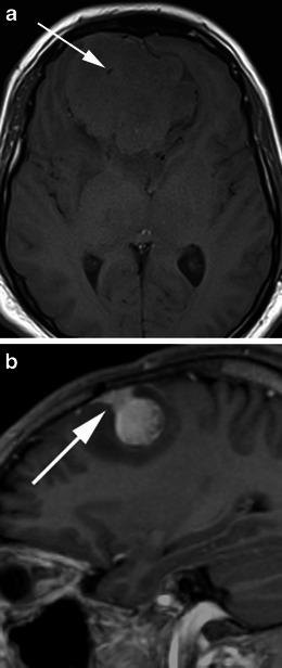
Fig. 19.

a Axial T1-weighted sequence in a 48-year-old man with presumed meningioma on CT demonstrates a large, extra-axial mass in the anterior cranial fossa within the interhemispheric fissure resulting in marked indentation of the medial aspect of the frontal lobes. The mass is predominantly isointense to grey matter and a number of flow voids are visualized around the periphery of the mass and centrally (arrow) in keeping with prominent vascularity. Histology at surgery was a haemangiopericytoma. b Post-contrast sagittal T1-weighted sequence in a 32-year-old man with a history of seizure post fall demonstrates an avidly enhancing, mildly heterogeneous, extra-axial mass indenting the right frontoparietal junction superiorly with a prominent pedicle (arrow) attached to the dura. A peripheral low signal intensity rim represents surrounding cortical grey matter and mild vasogenic oedema. Histology at surgery was a haemangiopericytoma
