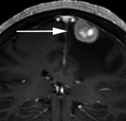
Fig. 20.

Post-contrast coronal T1-weighted sequence in a 70-year-old woman with headaches demonstrates a mildly heterogeneously enhancing mass indenting the right precentral gyrus (arrow) with a broad dural attachment and marked surrounding vasogenic oedema. The lesion was hyperintense to grey matter on the T2-weighted sequence. Histology at surgery was adenocarcinoma consistent with a breast primary
