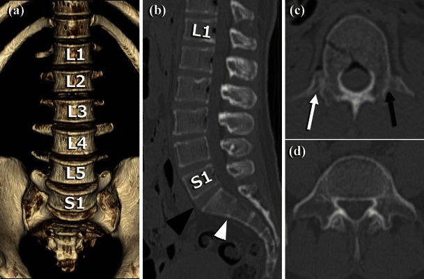
Fig. 4.

CT images of lumbarisation and unilateral lumbar rib. a VR image of the anterior aspect of the lumbar vertebrae showing lumbarised S1 segment. b Sagittal MPR image of the lumbar vertebrae showing a lumbar-type disk at S1-S2 (black arrowhead) and a sacrum-type disk at S2-S3 (white arrowhead). c Axial MPR image of L1 vertebra showing an articulated lumbar rib on the right side (white arrow) and a non-articulated transverse process on the left side (black arrow). d Axial MPR image of S1 with transverse processes, resembling L5
