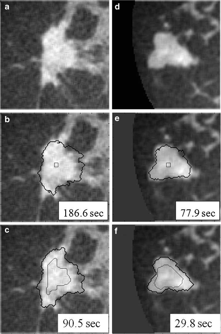
Fig. 2.
Comparison of active contour segmentation with different initial contours. a, d Coronal views of two dedicated breast CT lesions; b, e The initial contour was a cubic surface of 33 voxels; c, f The initial contour was, as included in our proposed overall segmentation method, an eroded RGI segmentation. Thin lines: initial contour; thick lines: final segmentation

