
Figure 2. The majority of IL-10 expressing splenocytes are uninfected.
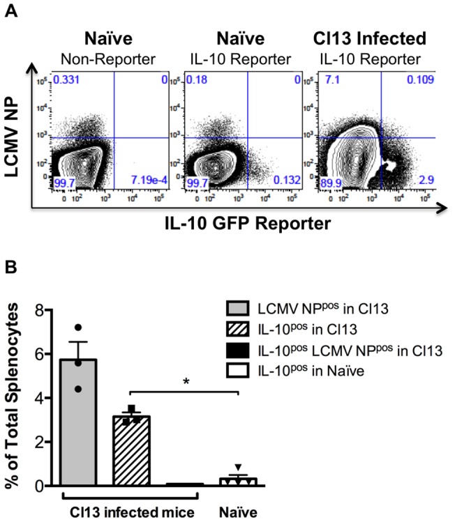
Adult IL-10 GFP reporter mice were infected with Cl13 and spleens harvested 15 days post infection. Splenocytes were stained with α-LCMV NP Ab. (A) Representative FACS plots are displayed. (B) The frequency of splenocytes that were LCMV NPpos (grey bar), IL-10 (GFPpos) (hatched bar), double positive (black bar) and the frequency of splenocytes that are IL-10pos in naïve reporter mice (white bar) is shown. n = 3 Cl13 and n = 4 for uninfected. SEM shown.