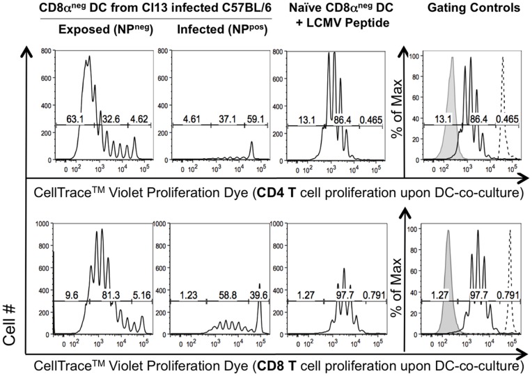
Figure 6. Directly infected CD8αneg DCs are unable to stimulate LCMV-specific CD4 and CD8 T cell proliferation.
DCs isolated from C57BL/6 mice infected in vivo with Cl13 7 days prior were sorted based on CD8α and LCMV NP surface expression. Sorted DCs were placed in culture with TCR transgenic LCMV specific CD4 T (Smarta) or CD8 T (P14) cells labeled with CellTraceTM Violet proliferation dye (CTV) and cultured for 4.5 days. Control cultures contained LCMV peptide (GP33 and GP61) pulsed CD8αneg DCs from naïve mice. Shown in the black histogram is the relative proliferation of the co-cultured Teff cells (gated on viability, CD4/8 T cell and congenic marker expression). Dashed line histograms are undivided CTV labeled Teff cells cultured alone, filled grey histograms are unlabeled cells. Each condition was set up in triplicate. Representative data from one of three independent experiments are shown.

