
Fig. 5.
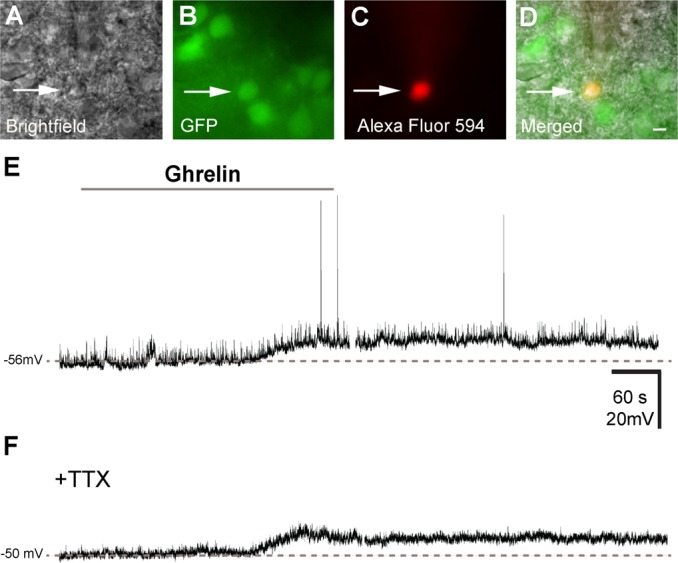
Ghrelin depolarizes a subset of Kiss1 neurons of the ARC. A: bright-field illumination of Kiss1-Cre/GFP neuron during acquisition of a whole recording (arrow). B: the same neuron under fluorescent (FITC) illumination to identify Kiss1-Cre/GFP signal. C: complete dialysis of Alexa Fluor 594 from the intracellular pipette at the end of the recording. D: colocalization of Alexa Fluor 594 and GFP in the same neuron. E: current clamp recording demonstrates that ghrelin (100 nM) depolarizes some Kiss1-Cre/GFP neurons. Dashed line indicates the resting membrane potential. F: current clamp recording demonstrates that ghrelin depolarizes Kiss1-Cre/GFP neurons in the presence of tetrodotoxin (TTX; 1 mM). Scale bar, 10 μm.