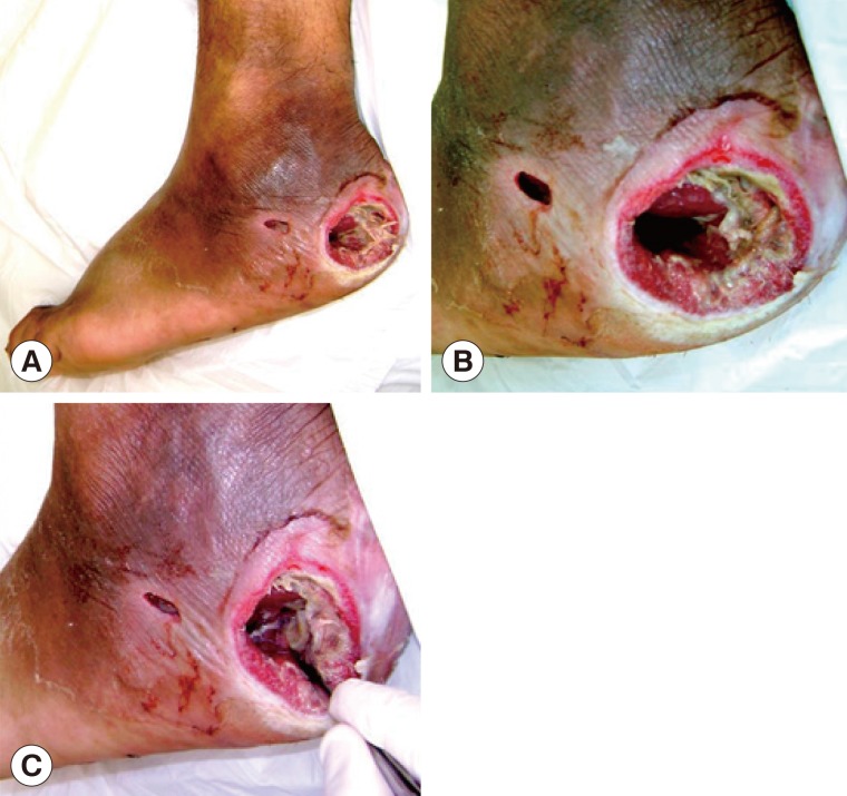
Fig. 1.
Diabetic foot ulcer region. (A) Clinical appearance of the ulcer in the heel of the right foot with the larvae inside it. (B) Cavitary lesion/ulcer caused by the larvae of Cochliomyia hominivorax. Note that the circular border is swollen and necrotic tissues are observed inside. (C) Mechanical extraction of the C. hominivorax larvae.

