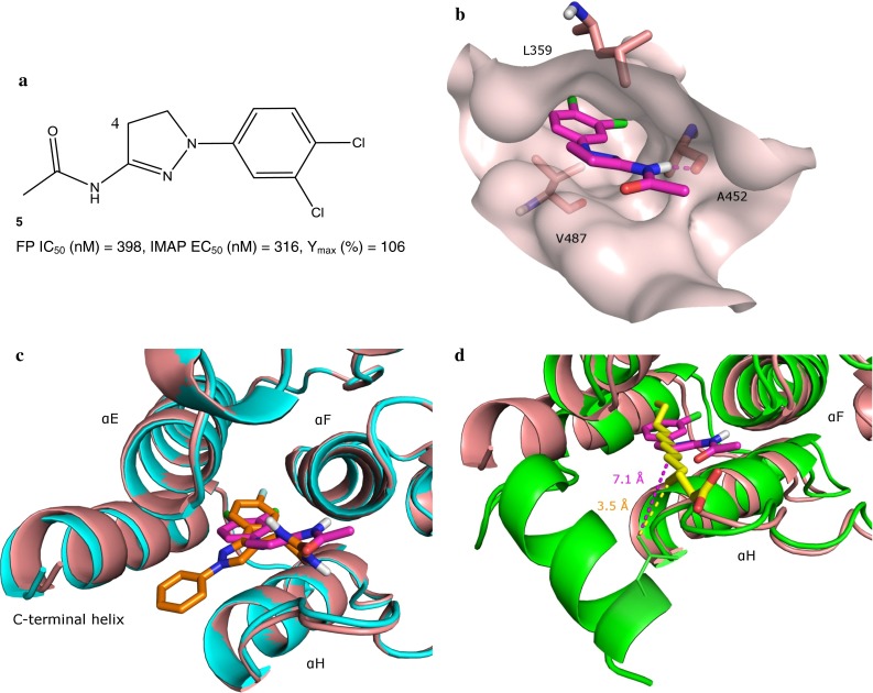
Fig. 3.

a Structure of dihydropyrazole exemplar 5 shown with its FP IC50 and IMAP EC50 values. Position 4 on the dihydropyrazole core is highlighted. b Interactions of 5 and the c-Abl myristoyl site (surface shown). For visual simplicity, only selected amino acids that interact with 5 are shown, and they are shown in pink. The carbon atoms of 5 are in magenta. Hydrogen bonds between 5 and the myristoyl site are indicated with dash lines. c Overlay of the c-Abl myristoyl site complexed to 1R (shown in cyan) and 5. d Overlay of 5-bound and myristoyl-bound c-Abl kinase domains. The shortest distance between the alkyl chain of the myristoyl and the side chain of Leu529 is illustrated in a yellow dash line; while the shortest distance between a lipophilic atom of 5 and the side chain of Leu529 is shown in a magenta dash line
