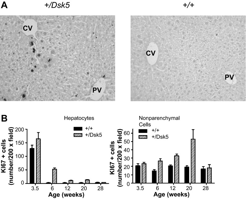
Fig. 2.
The hepatocytes and nonparenchymal cells of +/Dsk5 mice have a higher rate of cell proliferation. A: photomicrographs of liver sections from 6-wk-old male mice immunostained for Ki67, a cell proliferation marker. Note the nuclear staining in hepatocytes and the smaller nonparenchymal cells. The staining in hepatocytes tended to be greater around the central vein (CV), as opposed to the portal vein (PV). There were more proliferating cells in the +/Dsk5 mice; however, the number of proliferating cells was still much less than would be seen in early liver development or during liver regeneration following resection. B: quantification of Ki67+ cells obtained by counting the number of positively stained cells per ×20 field. The number of positive cells is shown as a function of age. Note that more nonparenchymal cells than hepatocytes stained for Ki67 after 6 wk of age in both +/+ and +/Dsk5 mice.

