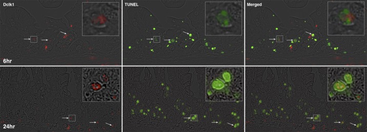
Fig. 2.

Identification of doublecortin-like kinase 1 (Dclk1)-positive apoptotic cells in small intestinal crypts after total body irradiation (TBI). The small intestine isolated 6 and 24 h post-TBI were fixed with formalin and embedded in paraffin. Paraffin-embedded sections were immunostained with anti-Dclk1 antibody (red on left) and ApopTag Peroxidase for apoptotic cells (green in middle). Colocalization of Dclk1 (red) in apoptotic cells (green) is indicated by arrows in the overlay pictures (right). Panels on top are 6 h post-TBI and bottom are 24 h post-TBI. The magnification is ×200, and the magnification for the inset is approximately ×600.
