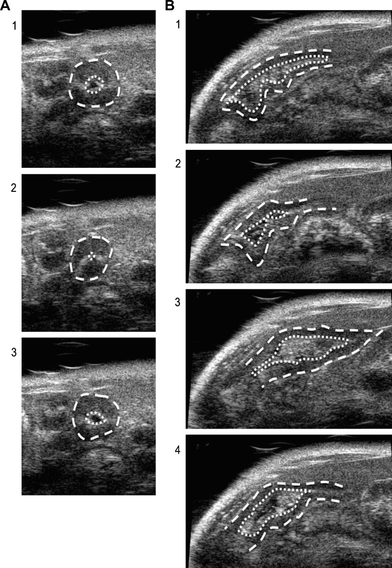
Fig. 4.
Ultrasound assessment of intestinal contraction. Sequential frames of a 30-frame/s video loop are shown for an axial (A, 1–3) and a longitudinal (B, 1–4) cross section of the intestine seen by ultrasound. Outlines of outer intestinal wall (dashed) and intestinal lumen (dotted) are indicated. A single contraction is depicted in each orientation from relaxed (1), to contracted with narrowing of the lumen and change of size/shape of intestinal wall (2), to relaxed as intestine reverts to original position (3–4).

