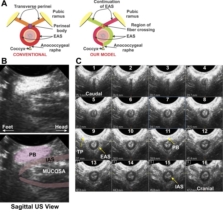
Fig. 1.
Schematic of our hypothesis (A) and sagittal and transverse ultrasound (US) images of the anal canal (B and C). A: schematic of conventional external anal sphincter (EAS) muscle architecture compared with our proposed model. B: sagittal US images of the anal canal. Note elliptical shape of the perineal body (PB), which has a well-defined structure. C: 16 serial axial images of the anal canal, spaced 15 mm apart. Note circular configuration of the internal anal sphincter (IAS), while the EAS merges with the transverse perineal (TP)/bulbospongiosus (BS) muscle at the ventral end. Cranial-caudal extent of the EAS and TP/BS is similar, and the EAS and TP/BS are continuous at the ventral end in the PB.

