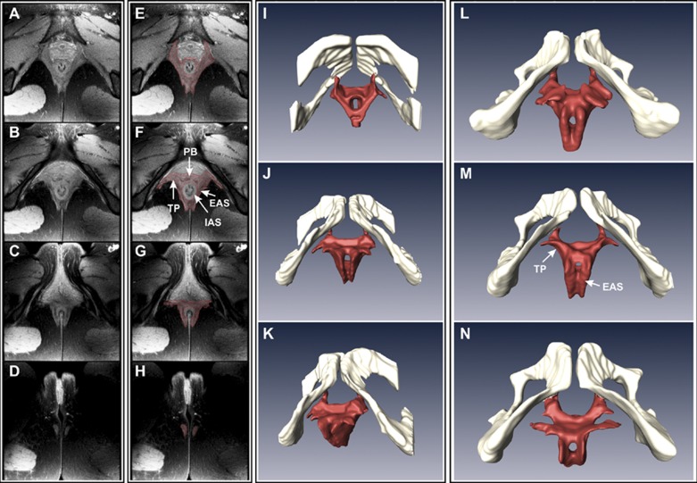
Fig. 2.
Proton density (PD) MRI of the anal canal (A–H) and 3-dimensional (3-D) reconstruction of the EAS muscle complex (I–N). In A–D, axial images of the anal canal (6 mm apart, from cranial to caudal direction) show various components of the anal sphincter complex. In E–H, muscle margins were marked manually to construct the 3-D anatomy (viewed from caudal to cranial direction, in 4 different subjects, I–N). I–K: images from 1 subject shown in 3 different projections. L–N: anatomy of the EAS muscle in the caudal-cranial projection from 3 additional subjects.

