Fig. 6.
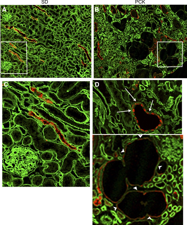
Laminin-511 is absent in the basement membranes of PCK cysts. Frozen sections of adult PCK rat kidneys and SD rat kidneys were double stained with a rabbit antiserum to laminin-α5 and a goat polyclonal antibody to AQP2 (red). The green color shows laminin-α5 staining. A: confocal image of a SD rat kidney section immunostained with laminin-α5 andAQP2 antibodies. There was uniform intensity of the green laminin-α5 staining in all tubular segments in this image. B: portion of a PCK kidney section with few cysts and precystic collecting ducts marked by AQP2 (red). Note that the green laminin-α5 staining was uneven, comparatively more in some tubular regions than in others, in the PCK kidney. C: magnified image of the region of SD rat kidney section enclosed by the white rectangle in A demonstrating the uniform distribution of laminin-511 in the collecting ducts (marked by red AQP2 staining) and other tubular regions not marked by AQP2 staining. D: two magnified images of PCK rat kidney sections showing the absence of laminin-511 (green) in the pericystic basement membrane (arrowheads) and in the basement membrane of precystic tubule (arrows).

