Fig. 1.

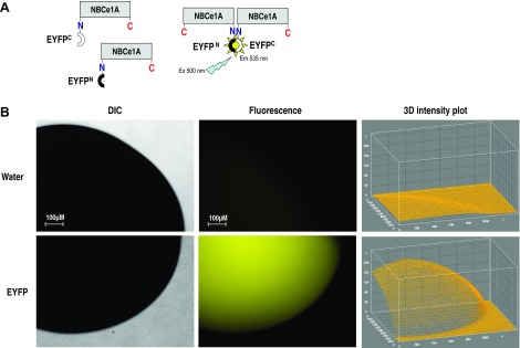
A: schematic illustration of biomolecular fluorescence complementation (BiFC) method used to visualize Na+/HCO3− cotransporter (NBCe1A) dimerization. Two complementary nonfluorescent fragments of the enhanced yellow fluorescent protein (EYFP) were ligated to the NH2 or COOH terminus of NBCe1A. Fluorescent signal reflects the dimer formation when we coexpressing YFPN-NBCe1A and YFPC-NBCe1A in the oocytes. We can mix match the YFPN and YFPC labeling linked to both NH2 termini or COOH termini or on either side to interpret the dimer folding is tail to tail, head to head, or head to tail fashion. B: full-length EYFP-pGEMHE construct expressed in Xenopus oocytes. Strong fluorescent signal from an unaltered EYFP protein in the expressing system verify the integrity of the EYFP protein. Minimum auto fluorescent signal was observed in the water-inject oocytes. The fluorescence intensity measured with ImageJ software was plotted on the Z axis of 3D intensity plot. DIC, differential interference contrast.
