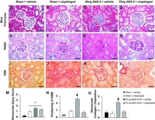
Fig. 6.

A–L: representative images of glomeruli from the four experimental groups stained with Masson's blue trichrome (A–D), periodic acid-Schiff-hematoxylin (PASH; E–H), or picrosirius red (PSR; I–L). Bars = 50 μm. M–O: quantification of the glomerular injury score (M), degree of glomerular basement membrane (GBM) thickening (N), and degree of glomerular collagen type I deposition (O). ND, not detectable. *P < 0.05 vs. the sham + vehicle-treated group; #P < 0.05 vs. all other groups.
