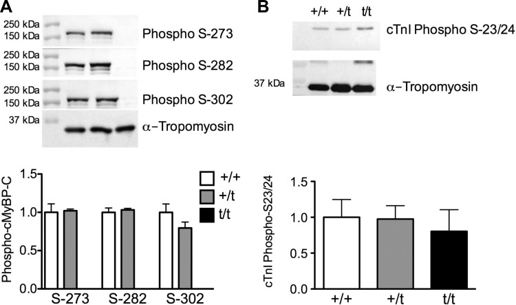
Fig. 2.
Phosphorylation of cMyBP-C and cTnI. A: Western blot analysis detecting cMyBP-C phosphorylated at serine-273, -282, and -302. No significant change in phosphorylation of cMyBP-C was observed at any of the phosphorylation sites between the +/+ and +/t groups (n = 5). B: phosphorylation status of cTnI at serine-23 and -24, as determined by Western blot, shows no significant changes in any of the groups (n = 5).

