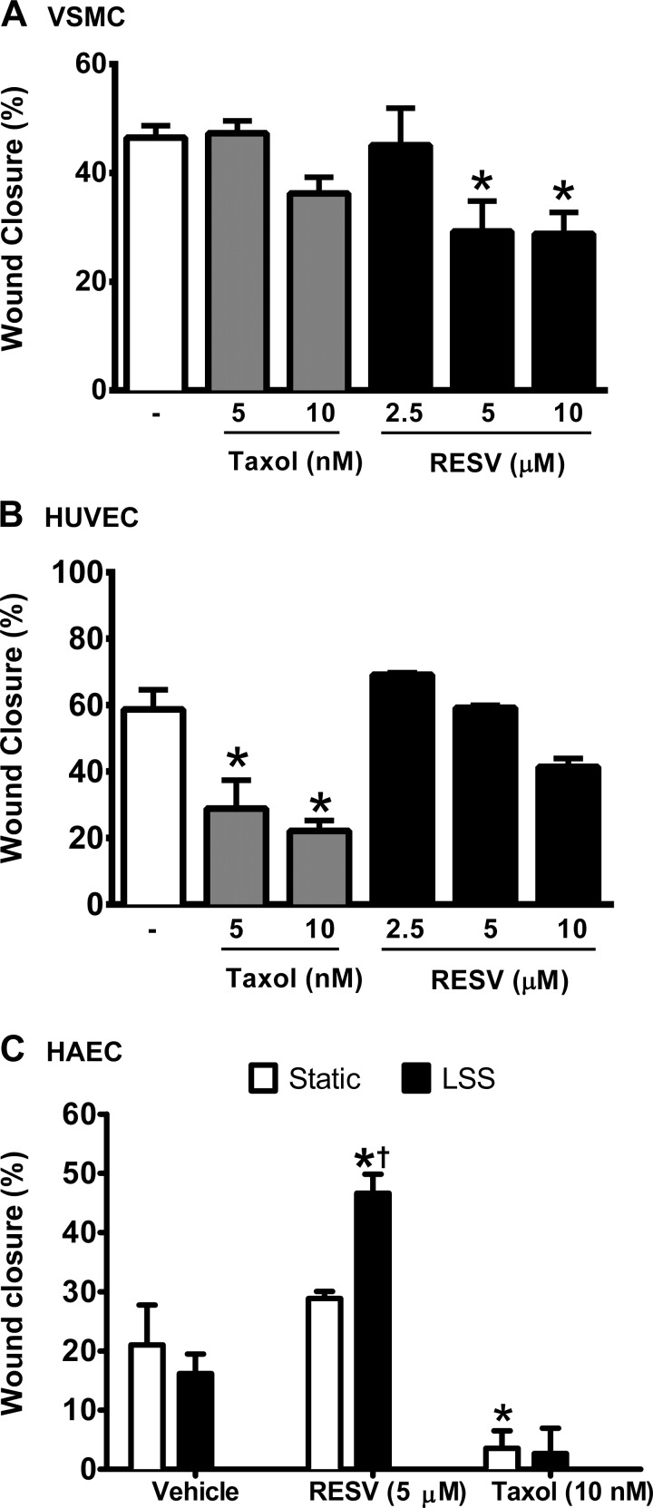
Fig. 2.
Taxol and RESV exert opposite selectivity in the vascular smooth muscle cell (VSMC) and endothelial cell (EC) wound healing assay. Wound closure was quantified in VSMC (A) compared with HUVEC (B), expressed as a percentage of the initial wound width that has healed. C: RESV and laminar sheer stress (LSS) interact to increase human aortic endothelial cell (HAEC) wound closure, quantified as the distance between the wound edges at 5 locations before and after injury. Data are means ± SE. *P < 0.05, compared with the appropriate vehicle control. †P < 0.05, compared with RESV-treated cells under static culture; n = 3–4.

