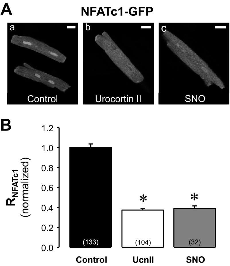
Fig. 3.
Effect of exogenous NO on subcellular NFATc1 distribution. A: confocal images of NFATc1-GFP distribution in control (a), UcnII (b), and spermine NONOate (SNO; c)-treated ventricular myocytes. B: average normalized RNFATc1 data in control, UcnII, and SNO. *P < 0.001 vs. control. Scale bar = 20 μm.

