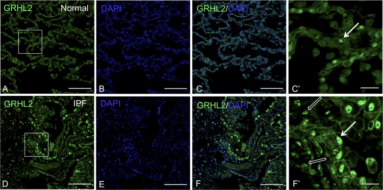
Fig. 2.
GRHL2 expression is observed in both lung epithelium and mesenchyme of idiopathic pulmonary fibrosis (IPF) lung. GRHL2 Immunofluorescence staining in normal (A–C) and IPF lung (D–F). C′ and F′ are insets of their respective images that lack DAPI for clarity. White arrows in C′ and F′ indicate epithelial cells whereas open white arrows in F′ indicate mesenchyme. Scale bar for A–F is 100 μm and for C′ and F′ it is 20 μm.

