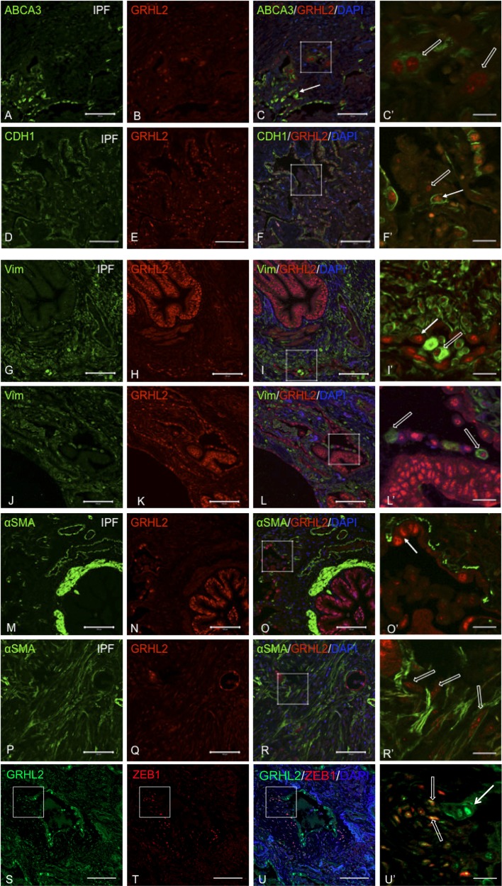
Fig. 4.
Colocalization of GRHL2 with epithelial and mesenchymal markers in IPF lung suggests cell-flattening events. Double staining of GRHL2 and ABCA3 (A–F) or CDH1 (G–I) or Vimentin (VIM; J–I) or α-smooth muscle actin (α-SMA; M–R) or ZEB1 (S–U). C′, F′, I′, L′, O′, R′, and U′ are insets of their respective images. White arrow in F, I′, L′, and O′ indicates epithelial nature of cells whereas open arrow in C′, F′, and I′ indicates cell flattening in collapsing alveolar regions. In R′, open arrows show myofibroblasts distant from collapsing alveoli. Scale bar for A–U is 100 μm and for C′, F′, I′, L′, O′, R′, and U′ it is 20 μm.

