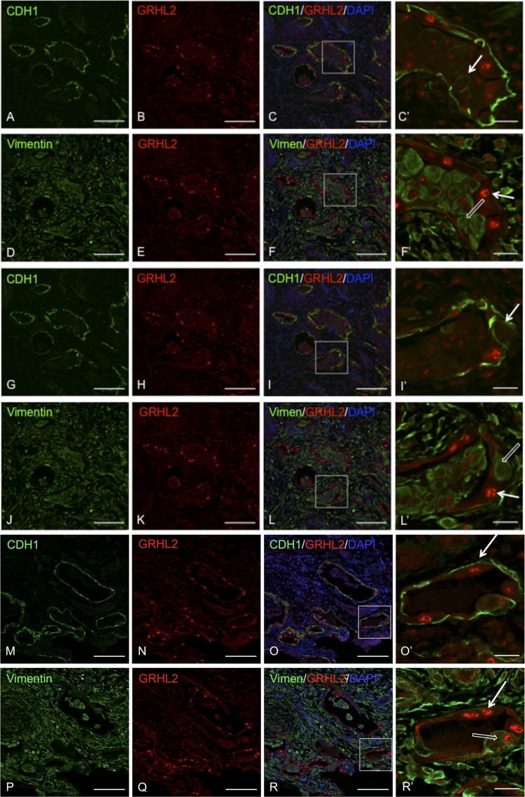
Fig. 5.
Colocalizaton of GRHL2 with epithelial and mesenchymal markers in serial sections shows cell-flattening events. Double staining of GRHL2 and CDH1 (A–C, G–I, and M–O). Double staining of GRHL2 and VIM in IPF lung is shown in (D–F, J–L, and P–R. C′, F′, L′, O′, and R′ are insets of their respective images. In C′, I′, and O′ white arrow indicates a subtype of epithelial cells that show bright GRHL2 and CDH1 signals, whereas in F′, L′, and R′ white open arrow indicates another subtype that has both CDH1 and VIM signals. Insets do not show DAPI for clarity. Scale bar for A–R is 100 μm and for C′, F′, I′, O′, and R′ it is 20 μm.

