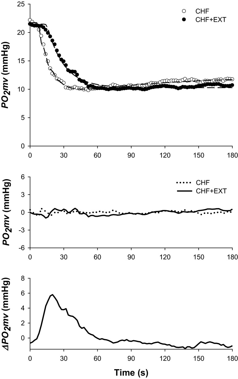
Fig. 1.
Top: Spinotrapezius muscle PO2mv profiles (circles) and their model fits (dashed lines) from representative chronic heart failure (CHF) and CHF + endurance exercise trained (EXT) rats in the control condition. Note that exercise training slowed the overall speed of the PO2mv fall during contractions. Middle: PO2mv residuals demonstrate excellent model fits. Bottom: absolute PO2mv difference between CHF + EXT and CHF responses shown at top. Note that the greatest effect of exercise training on PO2mv kinetics occurred during the rest-contraction transient (i.e., 0–60 s). Time zero denotes the onset of muscle contractions.

