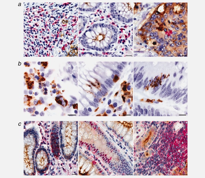
Figure 3.
COLCA1 and COLCA2 expression in paraffin-embedded colon biopsy samples. (a) Double immunohistochemical staining for COLCA1 (red; hematoxylin counterstain) and tumor-specific CEA (carcinoembryonic antigen; brown), scale bars, 20 µm. (b) 100× oil objective images (scale bars, 10 µm) of representative tissues immunostained with COLCA1 (brown; hematoxylin counterstain). (c) Double immunohistochemical staining for COLCA2 (red; hematoxylin counterstain) and CEA (brown), scale bars, 20 µm.

