
Figure 2. In vivo biodistribution after systemic administration of MV-m-uPA.
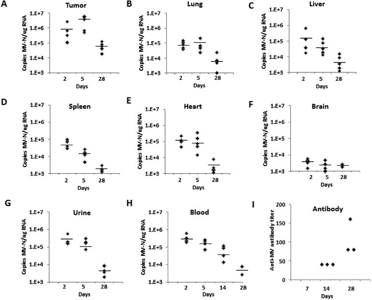
The orthotopic 4T1 tumor model was established in immunocompetent female Balb/c mice. Animals were treated and tissues processed as described in the methods section (n=5 mice per time point). At days 2, 5 and 28 days post-treatment, total RNA was extracted from frozen tumors (A), organs (B-F) and urine (G) for MV-N mRNA quantification by qRT-PCR. H. Blood samples were obtained for MV-N RNA quantification at days 2, 5, 14 and 28 after treatment (n=5 per time point). Results were expressed as copies of MV-RNA/μg of total RNA in each organ/tissue, and horizontal bars represent the mean value of the replicates. (I) Determination of serum anti-MV antibody. Serum was obtained from treated mice at 7 (no antibody detected), 14 and 28 days after treatment (n=3 per time point) for antibody determination (see methods section for details).