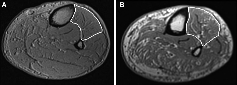
Fig. 1.
Sample MRI images of age-matched (∼65 yr old) male control (A) and diabetic polyneuropathy (DPN) patient (B) leg. The tibialis anterior (TA) is outlined in white. Note the greater amounts of intramuscular fatty infiltration and noncontractile tissue found in the DPN patient leg compared with the control leg (also see Table 3).

