Fig. 5.

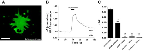
Cells stained with the reduced HAF protocol consistently report an alkalinization when challenged with kainate or glutamate. A: epifluorescent (nonconfocal) derived image of an isolated catfish horizontal cell exhibiting membrane-associated HAF staining via revised 500 nM HAF staining protocol. Scale bar, 25 μm. B: representative response from 1 cell exhibiting robust membrane-associated staining and challenged with 50 μM kainic acid; an extracellular alkalinization (increase in fluorescent ratio) is observed. C: graph showing the peak amplitude fluorescence change when challenged with 50 μM kainate or 300 μM glutamate (black bars); the extracellular alkalinization was blocked when kainate was delivered in the presence of CNQX, nifedipine, or lanthanum (gray bars). *Statistically significant difference between the response to 50 μM kainate and 300 μM glutamate; **statistically significant difference in presence of blockers compared with responses induced by kainate or glutamate.
