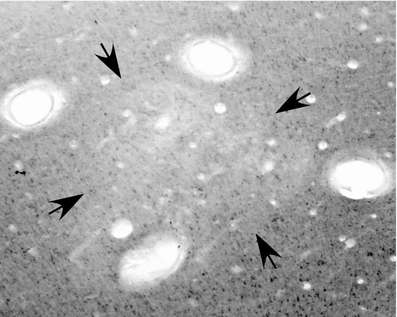
Fig. 1.
A cortical section from PMD-M1 border region cut parallel to the surface and stained for cytochrome oxidase (CO) in G 08–01. Note the reduced CO level within the area marked by arrows, and the four electrolytic lesions (open ovals) placed at the functional borders of the reach domain. Muscimol was injected at three sites within the domain (see Fig. 7B).

