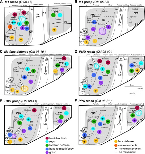
Fig. 14.
Summary results of inactivation of functional domains in frontal (M1, PMD, and PMV) and posterior parietal (PPC) cortex. Inactivation of reach and grasp domains in M1 (A and B), face domain in M1 (C), the PMD reach domain (D), the PMV grasp domain (E), and the PPC reach domain (F). Hamilton syringes depict the approximate spatial center of the muscimol injections and the surrounding circle with dots depicts the inactivated territory. Presence or lack of movements after cortical inactivation are indicated by “+” or “-” shown on color circles corresponding to particular movement domains (see Figs. 2 and 3). Solid lines mark areal borders, and dashed lines mark borders between body representations. Other conventions as in Figs. 2 and 3.

