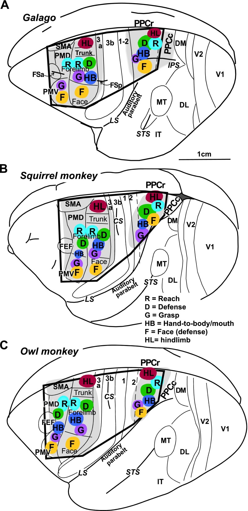
Fig. 2.
Schematic illustration of functional domains in frontal and parietal cortex in the left hemisphere of prosimian galago (A), squirrel monkey (B), and owl monkey (C). Territories investigated with microstimulation are depicted in light grey and the approximate locations of functional domains are depicted in color. The black frame corresponds to outlines of schematics in summary Fig. 14. Motor cortical areas: FEF, frontal eye field; M1, primary motor; PMD, dorsal premotor; PMV, ventral premotor; SMA, supplementary motor. Somatosensory cortical areas: 3a, 3b, 1–2. Visual cortical areas: DL, dorsolateral; DM, dorsomedial; FST, fundal superior temporal; IT, inferotemporal; MT, middle temporal; MTc, middle temporal crescent; MST, middle superior temporal; V1, primary visual; V2, secondary visual; V3, third visual. Sulci: CS, central sulcus; FSa, anterior frontal; FSp, posterior frontal; IPS, intraparietal; LS, lateral; STS, superior temporal; PPCr, rostral region of the posterior parietal cortex.

