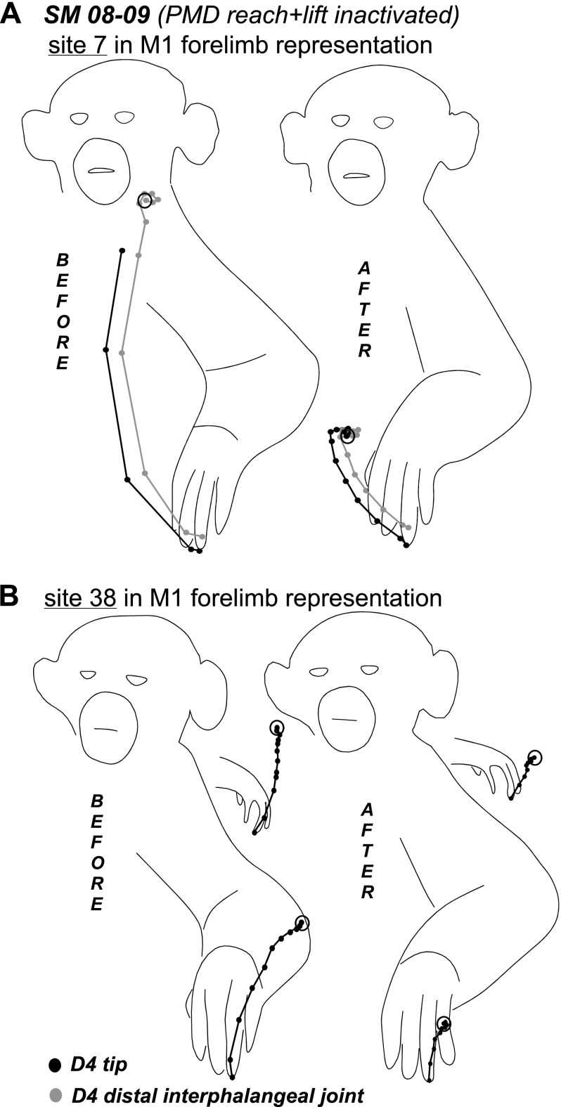
Fig. 8.
Trajectories of hand-to mouth (A) and lateral reach movements (B) evoked from sites 7 and 38, respectively, in the M1 forelimb representation of SM 08–09. Trajectories show the original movement and the movement evoked during inactivation of the PMD reach domain. Black dots indicate the positions of the tip of digit 4 and gray dots indicate the position of distal interphalengeal joint in successive video frames. Note the truncated trajectories of both movements evoked after muscimol inactivation. For site 38, trajectories of hand movements reflected in the mirror are also shown. Other conventions as in Fig. 6.

