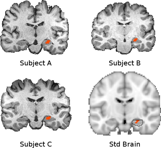
Fig. 4.
The hippocampal seed shown in standard space (bottom right, orange, Y = −14) and native space for 3 subjects. The region of interest (ROI) was defined in standard space according to a region that showed greater connectivity extent in pain patients compared with CON subjects. This region is circumscribed by the hippocampus defined in the Harvard-Oxford subcortical atlas (bottom right, black outline), which identifies the likelihood of any particular voxel belonging to a particular subcortical structure (black outline indicates P > 0.5 likelihood for hippocampus). This ROI was transformed into native space for seed-based analysis in each subject. The fidelity of this transformation is indicated with T1 volumes from 3 representative subjects with the corresponding native space seed overlaid. Connectivity maps were transformed back into standard space subsequently for higher-level analysis.

