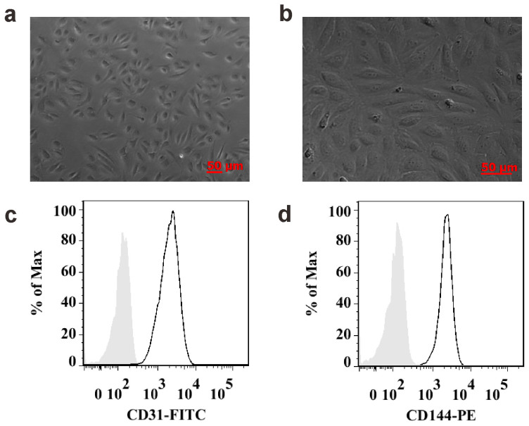
Figure 2. Identification and characterization of HUVECs.
(a and b) Microscopic morphology: exhibited typical cobblestone growth pattern of endothelial cells (a), HUVECs become tightly packed but show no tendency to overlap or overgrow one another (b). (c and d) HUVECs purity was assessed by flow cytometry: CD31, 99.5%; and CD144, 99.6%, compared with isotype control (silver area).

