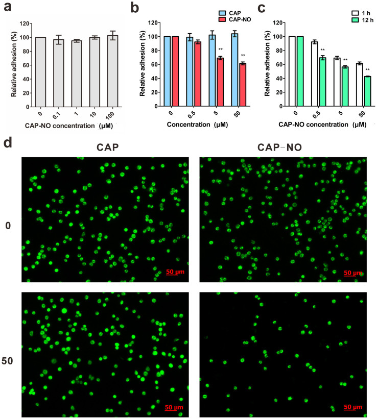
Figure 4. CAP-NO inhibited adhesion of HT-29 cells to HUVECs.
(a) Effect of CAP-NO on the spontaneous adhesion of HT-29 cells to culture plate within 1 h observation. (b) Quantification of HT-29 adhered to the HUVEC monolayers in the presence and absence of CAP-NO or CAP. (c) Effects of CAP-NO on adhesion of HT-29 to HUVECs at indicated time points. The % relative adhesion was determined by fluorescence-labeled cell count assay, and the results are based on the IL-1β stimulated HUVECs. (d) Rhodamine 123-labeled HT-29 cells were added to the HUVEC monolayers stimulated by IL-1β (1 ng/mL) in the presence and absence of CAP-NO or CAP (both 50 μM). Fluorescence microscopy showed HT-29 cells (green) adhered to the HUVECs. Bars represent the mean ± SEM (n = 3); ** indicates P < 0.01.

