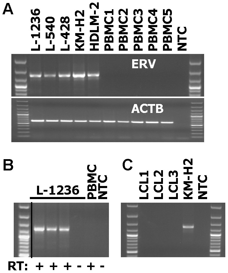
Figure 3. Expression of ERVK_1q42.13 in HL cell lines.

(A) Presented are results from a RT-PCR with primers with specificity for actin beta (ACTB) and ERVK_1q42.13. cDNA from HL cell lines and normal PBMC was used as template for PCR. NTC: no template control. (B) Presented are results from a RT-PCR with primers with specificity for ERVK_1q42.13. cDNA from HL cell line L-1236 and normal PBMC was used as template for PCR. cDNA was synthesized by using reverse transcriptase (RT:+) from three different vendors (see Material and methods). In addition, RNA was used without reverse transcription (RT:-). NTC: no template control. The black dividing line indicates removal of five irrelevant lanes from the original image. (C) Presented are results from a RT-PCR with primers with specificity for ERVK_1q42.13. cDNA from HL cell line KM-H2 and three different lymphoblastoid cell lines (LCL) was used as template for PCR. NTC: no template control.
