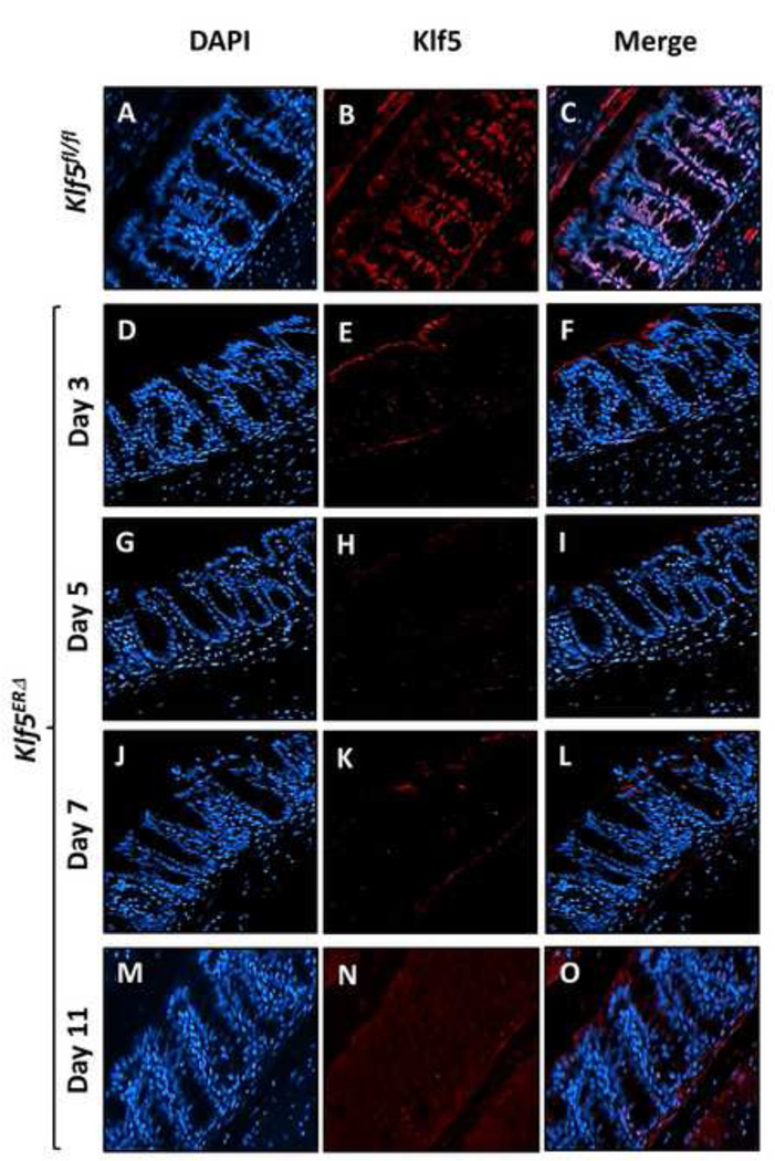
Figure 2. Tamoxifen treatment of Klf5ERΔ mice results in deletion of Klf5 from colonic tissues.
Immunofluorescence analysis of Klf5 expression in colonic tissues from both Klf5ERΔ and control Klf5fl/fl mice after tamoxifen treatment. Images are stained with DAPI (blue), Klf5 (red) and merged (DAPI + Klf5). Panels A through C show colonic tissues from day 5 tamoxifen-treated control Klf5fl/fl and Panels D through O, those from Klf5ERΔ mice. Panels D, E & F are representative of 3 day tamoxifen treatment of Klf5ERΔ mice. Panels G-I, J-L and M-O represent 5, 7, and 11 day post-tamoxifen treatment respectively. Klf5ERΔ mice show a complete loss of Klf5 (Panels E, H, K & N) from the nuclei of colonic epithelial cells when compared to Klf5fl/fl colon (Panel B).

