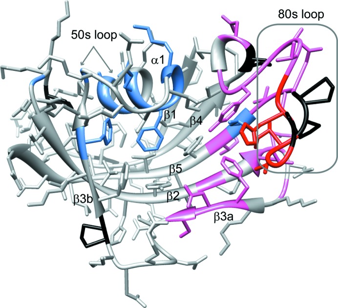
Figure 5.
Structural distribution of residues exhibiting amide-resonance doubling owing to slow conformational exchange in FKBP12.6. Residues that yield doublings of their amide resonances separated by more than 0.15 p.p.m. (averaged as Δ1H and 0.2Δ15N; Garrett et al., 1997 ▶) are indicated in red. Residues exhibiting smaller chemical shift differences between the two conformational states are indicated in pink. Residues that exhibit doubling in FKBP12 (Mustafi et al., 2013 ▶) but not in FKBP12.6 are indicated in blue. Prolines are marked in black.

