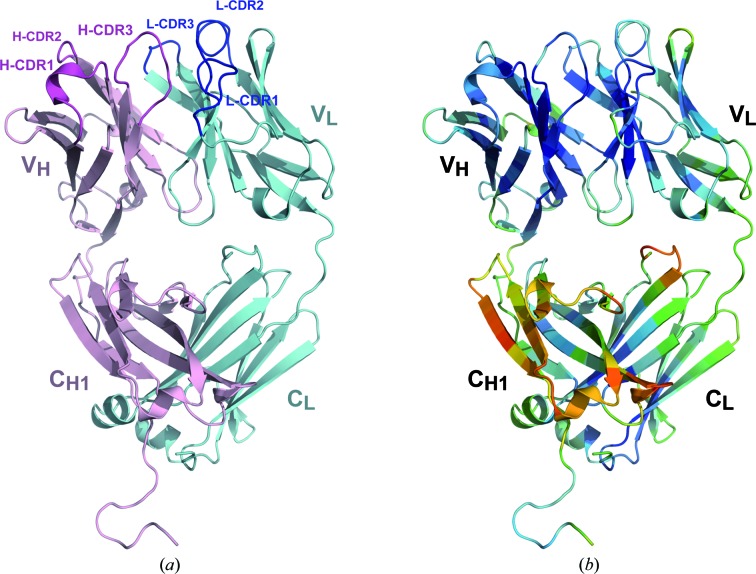
Figure 2.
(a) The overall structure of the A17λ antibody. Heavy (VH/CH1) and light chains are shown in magenta and cyan, respectively. (b) The A17λ structure coloured according to the Cα atomic displacement parameters (ADP), with a colour transition from blue to red indicating increasing ADP values.

