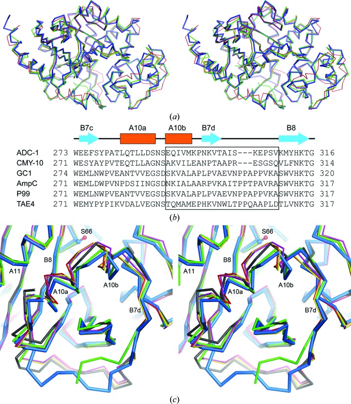
Figure 7.
Comparison of ADC-1 with other class C β-lactamases. (a) Stereoview of the superposition of ADC-1 (thick blue ribbon) on CMY-10 (green ribbon) and AmpC (red ribbon). (b) Structure-based sequence alignment of the R2-loop region of ADC-1 with five representative class C β-lactamases: CMY-10, GC1, E. coli AmpC, P99 and TAE4. The R2-loop residues are indicated by the black box. (c) Stereoview of the superposition of ADC-1 (blue ribbon) with the same five class C β-lactamases as in (b): CMY-10 (green), GC1 (magenta), AmpC (red), P99 (yellow) and TAE4 (black). The R2-loop extends from the beginning of helix A10b to the beginning of strand B8.

