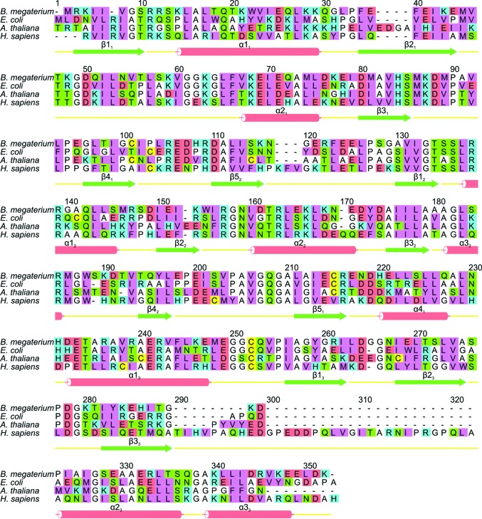
Figure 4.
Sequence alignment and secondary structure of B. megaterium PBGD. An alignment of B. megaterium PBGD with the enzyme from another prokaryote (E. coli) along with the plant (A. thaliana) and human enzymes. The secondary-structure elements are labelled using the notation of Louie et al. (1992 ▶) and the amino-acid residues are colour-coded as follows: cyan, basic; red, acidic; green, neutral polar; pink, bulky hydrophobic; white, Gly, Ala and Pro; yellow, Cys.

