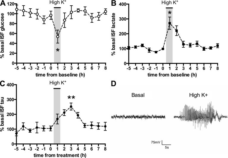
Figure 1.
Depolarization increases tau in ISF. (A–C) Microdialysis experiments were performed in hippocampi of wild-type mice. After baseline collection, the regular perfusion buffer was switched to high K+ perfusion buffer (administration indicated by gray box). After 1 h, the buffer was switched back to normal perfusion buffer (wash out) and ISF collection was continued. Glucose (A), lactate (B), and tau (C) in ISF were measured. (n = 5; *, P < 0.05; **, P < 0.01). For mice studied in A–C, each mouse was investigated independently. Any treatment effects were compared with baseline values within each mouse. Error bars represent SEM. (D) Representative EEG trace from one of three mice during depolarization with high K+ perfusion buffer.

