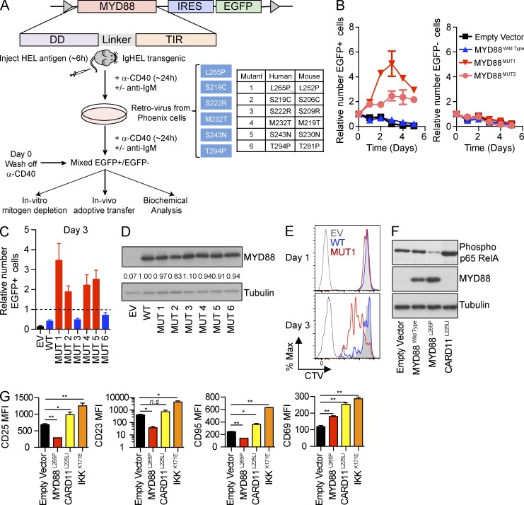
Figure 1.
MYD88 mutations promote mitogen-independent B cell proliferation in vitro but, paradoxically, shut down NF-κB. (A) Experimental strategy for retrovirally introducing mutant MYD88 into activated mature splenic B cells. HEL-specific B cells from IgHEL-transgenic mice were activated by HEL antigen in vivo and polyclonal B cells from nontransgenic mice were activated by including anti-IgM during culture with anti-CD40. After transduction, the cells were washed and cultured without these mitogens (day 0) or transplanted into syngeneic mice. Table shows the corresponding human and mouse amino acid substitutions tested. (B) Activated HEL antigen-specific B cells transduced with the indicated vectors were placed into fresh triplicate cultures in the absence of antigen or CD40 stimuli for 5 d. Mean and SD of EGFP+ cells expressing each vector were compared with the starting amount on day 0 of the cultures. Data are representative of at least three independent experiments. (C) Mean and SD of EGFP+ B cells transduced with the indicated vectors after 3 d in triplicate cultures without antigen or CD40 stimulation, relative to the number at the start of the cultures (dashed line) are representative of at least three independent experiments. (D) EGFP+ cells expressing the indicated vectors were sorted on day 1 of culture without antigen or CD40 stimuli, and lysates analyzed by SDS-PAGE and Western blot for MYD88 protein and tubulin as loading control. Numbers show densitometric measurements of MYD88 in each sample, expressed as relative to cells expressing the MYD88WT vector. (E) Cell division measured by CTV dilution on days 1 and 3 of culture without antigen or CD40 stimulation, gated on MYD88L265P:EGFP (MUT1) or MYD88WT:EGFP-transduced (WT) B cells or in nondividing empty:EGFP vector transduced B cells (EV). Unlabeled cells are shown by the open gray histogram. Data are representative of at least three independent experiments. (F) EGFP+ cells expressing the indicated vectors were sorted on day 1 of culture without antigen or CD40 stimuli, and lysates were analyzed by SDS-PAGE and Western blot for phosphorylation of the p65 subunit of NF-κB, with the blot sequentially reprobed with antibodies to MYD88. Tubulin was used as a loading control. (G) Mean fluorescent intensity (MFI) of cell surface expression of proteins encoded by NF-κB–inducible genes, measured in independent triplicate cultures (mean and SD) by flow cytometry on day 1 of culture without antigen or CD40 stimuli, gated on EGFP+ cells transduced with the indicated vectors. Statistical analysis by Student’s t test. ns, not significant; *, P < 0.05; **, P < 0.01. Data are representative of at least three independent experiments.

