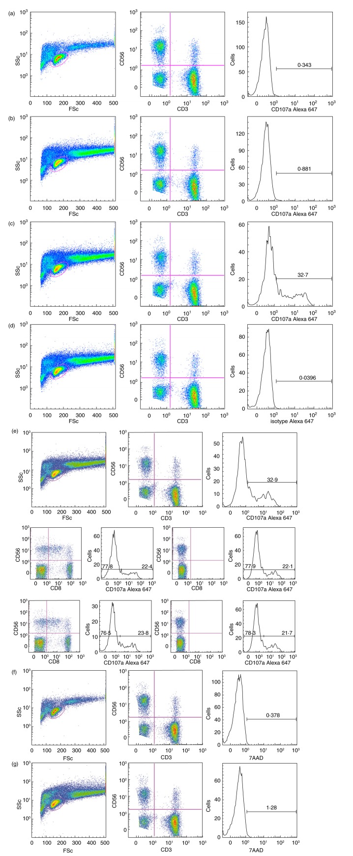
Figure 1.
Flow cytometry gating strategy and controls. Fluorescence activated cell sorter (FACS) profiles illustrating gating strategy and controls – in this case with effector A and the target cell culture MS1946. The samples are: a: effector A alone: from left to right left: ungated [side scatter (SSc) versus forward scatter (FSc]; centre: lymphocyte gate (CD56 versus CD3); right: CD3−CD56+ gate (CD107a); b: effector A with target cells: left: ungated (SSc versus FSc); centre: lymphocyte gate (CD56 versus CD3); right: CD3−CD56+ gate (CD107a); c: effector A with target cells and Rituximab®: left: ungated (SSc versus FSc); centre: lymphocyte gate (CD56 versus CD3); right: CD3−CD56+ gate (CD107a); d: effector A with target cells and Rituximab® (isotype control): left: ungated (SSc versus FSc); centre: lymphocyte gate (CD56 versus CD3); right: CD3−CD56+ gate (isotype control for CD107a); e: effector A with target cells and Rituximab® (FMO control): (e), top row: left: ungated (SSc versus FSc); centre: fluorescence-minus one (FMO)CD8 lymphocyte gate (CD56 versus CD3); right: CD3−CD56+ gate (CD107a); (e), continued, mid-row: far left: lymphocyte gate (CD56 versus CD8); left: CD56+ (includes CD56+CD8+ and CD56+CD8−)(CD107a); right: FMOCD8 lymphocyte gate (CD56 versus CD8); far right: FMOCD8 CD56+(CD107a); (e), continued, bottom row: far left: lymphocyte gate (CD56 versus CD8); left: CD56+ (CD56+CD8− only) (CD107a); right: FMOCD8 lymphocyte gate (CD56 versus CD8); far right: FMOCD8 CD56+ (CD107a). Note that (e), continued, mid-row and bottom row present the same analysis with two different gating strategies. (f) Effector A alone, left: ungated (SSc versus FSc); centre: lymphocyte gate (CD56 versus CD3); right: natural killer (NK) gate with the viability marker 7-aminoactinomycin D (7AAD) (CD3−CD56+) (7AAD); (g) effector A with target cells and Rituximab, left: ungated (SSc versus FSc); centre: lymphocyte gate (CD56 versus CD3); right: NK gate with the viability marker 7AAD (CD3−CD56+)(7AAD). Anti-CD8 antibodies were also added to all samples, except for (e) FMOCD8 controls (f,g): viability marker.

