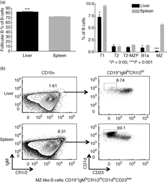
Figure 2.

B cell subsets in the liver and spleen. Single cell suspensions of liver and spleen were blocked for 10–15 min with anti-CD16/32 and Normal Rabbit Serum (NRS) and stained with dead cell dye aqua, followed by staining with a fluorescent-tagged antibody mixture directed against the cell surface markers CD1d, CD3, CD5, CD19, CD23, CD24, CR1/2, IgM and IgD. B cells were gated on CD19+ cells after exclusion of dead cells and CD3+ cells. (a) Six immature and mature B cell subsets were identified as follows: transitional T1 B cells are CD19+CD24hiIgMlowCD23-; transitional T2 cells are CD19+CD23hiIgMhiCR1/2low; transitional T2-marginal zone-precursor (T2-MZP) B cells are CD19+IgMhiCD23hiCR1/2hi; follicular B cells are CD19+IgMlowCD5-CD23hiIgDhiCR1/2low; MZ-like cells are CD19+IgMhiCD5-CD23lowIgDlowCR1/2hi CD1dhi; B1a B cells are CD19+immunoglobulin (Ig)MhiCD5+CD23lowIgDlowCR1/2low. B1b B cells that are CD11b+ were too few to quantify in the spleen and liver. Data are plotted as % of CD19+ B cells. The liver exhibits a greater proportion of follicular B cells and a smaller proportion of T1, B1a and especially marginal zone (MZ)-like cells; *P < 0·05; ***P < 0·001; n = 4 mice. Data are representative of two independent experiments. (b) Representative flow plots for MZ-like B cells. Left panels are gated on CD19+ cells. Right panels are gated on CD19+ IgMhiCR1/2hi.
