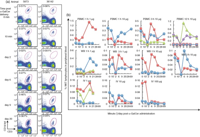
Figure 1.
Transient depletion of peripheral natural killer T cells (NKT cells) upon in-vivo α-galactosylceramide (α-GalCer) delivery. (a) Representative plots of flow cytometry analysis of pigtail macaque NKT cells within the lymphocyte population of peripheral blood stained ex vivo at different times following α-GalCer delivery; animal 5873 was administered 10 μg α-GalCer pulsed onto peripheral blood mononuclear cells (PBMC) for 12 h, animal 36142 was administered 1 μg α-GalCer pulsed onto WB for 3 h. NKT cell levels are enumerated as cells double-positive for CD1d tetramers loaded with the α-GalCer analogue, PBS-44, and CD3 as a proportion of gated lymphocytes. (b) Sequential blood samples were taken prior to delivery (0 min) and at 10 min, days 3, 9, 20 and 28 following 1 μg α-GalCer administration (n = 8) (b, left column) or at 0 min, 10 min, days 2, 6, 9, 20, 69, following 1–100 μg α-GalCer delivery (n = 19) (b, columns 2–4). Each line represents the peripheral NKT cell frequency of one animal. Dose and mode of administration of α-GalCer are indicated above each individual graph.

