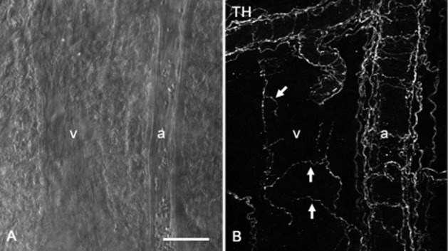
Figure 6.

Sympathetic innervations of submucosal microvasculatures of the rat distal colon. A venule (v) and an arteriole (a) running parallel were observed in the submucosal specimen using a differential interference contrast microscope (A). In the same specimen, TH-immunoreactive sympathetic varicose nerve fibres (arrows) were sparsely distributed around a submucosal venule (v), while bundles of sympathetic fibres ran along a submucosal arteriole (a) (B). Scale bar: 50 μm.
