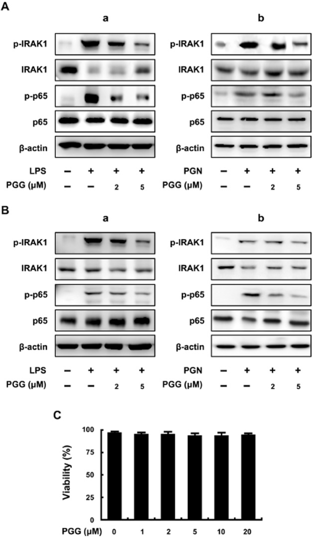
Figure 1.

Effect of PGG on IRAK1 phosphorylation and NF-κB activation in LPS- or PGN-stimulated peritoneal and colonic macrophages. (A) Effect in peritoneal macrophages stimulated with LPS (a) or PGN (b). (B) Effect in colonic macrophages stimulated with LPS (a) or PGN (b). Peritoneal macrophages (5 × 10⁵ cells) were treated with 50 ng·mL−1 LPS in the absence or presence of PGG (2 or 5 μM) for 20 h. IRAK1, p-IRAK1, p65, p-p65 and β-actin levels were measured by immunoblotting. (C) Cytotoxicity in peritoneal macrophages. Peritoneal macrophages (5 × 10⁵ cells) were treated with PGG (2, 5 and 20 μM for 48 h.
