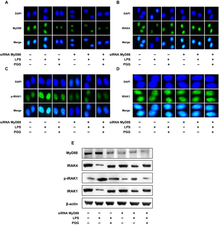
Figure 5.
Inhibitory effect of PGG on IRAK1 and NF-κB activation in LPS-stimulated peritoneal macrophages transfected with MyD88 siRNA. (A) Effect on MyD88 expression. (B) Effect on IRAK4 expression. (C) Effect on p-IRAK1 expression. (D) Effect on IRAK1 expression. Peritoneal macrophages isolated from mice were treated with 50 ng·mL−1 LPS in the absence or presence of PGG (5 μM) for 90 min. Alexa Fluor 488-conjugated IRAK1 and p-IRAK1 were detected in siRNA-transfected peritoneal macrophages. (E) Effect on IRAK1 and IRAK4 degradation and IRAK1 phosphorylation. MyD88, IRAK1, p-IRAK1, IRAK4 and β-actin were analysed by immunoblotting. Peritoneal macrophages were treated with 50 ng·mL−1 LPS in the absence or presence of PGG (5 μM) for 90 min.

