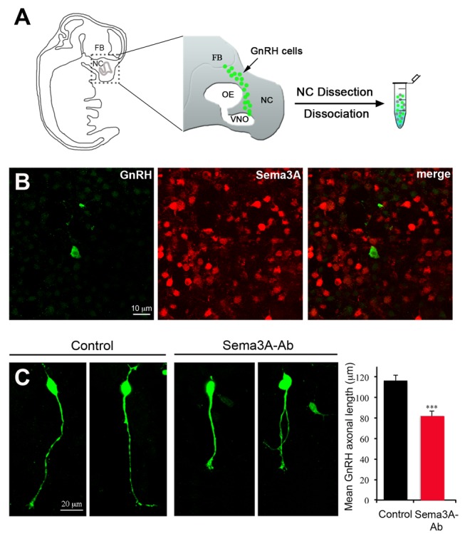
Figure 4. Sema3A immunoneutralization causes the retraction of axon-like processes in primary GnRH neurons in vitro.

(A) Schematic representation of a sagittal view of a mouse embryo at E12.5, showing the distribution of GnRH neurons (green dots) within the head. Primary cultures were performed from microdissected nasal compartment (NC) explants, which contain most GnRH neurons at this embryonic stage. FB, forebrain; OE, olfactory epithelium; VNO, vomeronasal organ. (B) Representative images showing the binding of the Sema3A-neutralizing antibody (red) to cultured cells surrounding GFP-expressing GnRH neurons (green). (C) Representative images showing the morphology of cultured GnRH neurons under control conditions and after treatment with the Sema3A-neutralizing antibody and bar graph quantifying the mean length of their axon-like processes (control, N = 3 independent experiments, n = 146 cells; Sema3A-Ab, N = 3 independent experiments, n = 143 cells). Total number of cultures, 24 from 4 litters. Data are represented as means ± SEM. Unpaired Student's t test, t (285) = 4.823, p<0.0001.
